

How Do Plant And Animal Cell Mitosis Differ
Mitosis in plants and animals
Microscopy - Teacher's Guide
SED 695B; Fall 2005


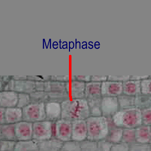
Onion root
Whitefish Blastula
Topics addressed
Clarification of Investigation
- Mitotic cycle
- The role of Chromosomes in heredity
- Comparing of plant and animal cells
Today you will use prepared slides to investigate the phases of mitosis in both plants and animals.
- Longitudinal sections of the root of Allium, will be used to examine the cells in the root meristem, the growing region of the root.
- Whole mounts of whitefish blastula will illustrate reproductive cells in animals. These undifferentiated cells undergo mitosis at a regular interval every bit the embryo increases in number of cells and complication.
- You volition make observational drawings and be prepared to take a practical quiz.
Standards:
High School:
Cell Biology 1. The fundamental life processes of plants and animals depend on a variety of chemical reactions that occur in specialized areas of the organism's cells. As a footing for understanding this concept:
c.
Students know how prokaryotic cells, eukaryotic cells (including those from plants and animals), and viruses differ in complication and general construction.
Center Schoolhouse:
Prison cell Biology 1. All living organisms are composed of cells, from simply one to many trillions, whose details usually are visible only through a microscope. As a ground for understanding this concept:
b.
Students know the characteristics that distinguish plant cells from fauna cells, including chloroplasts and prison cell walls. c.
Students know the nucleus is the repository for genetic information in plant and fauna cells. due east.
Students know cells split to increment their numbers through a process of mitosis, which results in two daughter cells with identical sets of chromosomes. f.
Students know that as multicellular organisms develop, their cells differentiate.
Study Guide:
Found and Animal Mitosis
Your objective:
Find and make observations of cells in each phase of mitosis in plant and creature tissue. Compare the differences between plant and animal mitosis. Be able to correctly identify the phases from both plant and animal tissue.
Materials:
- Prepared slide labeled 'Allium root, mitosis'
- Prepared slide labeled 'whitefish blastula, mitosis'
- compound microscope
Procedures:
- Prepare your microscope, identify the onion root slide on the phase and focus on low (40x) power.
- move your slide then that your field of view is centered on the root tip.
- Focus at 100x and re center so that you are focused on the more 'foursquare' meristem cells.
- Focus at 400x.
- Slowly move the slide and search for cells in each phase of mitosis
- When you have found one, make a detailed observational cartoon of that cell. Label all important structures (nucleus, chromosome, cell plate). Title the drawing with the blazon of prison cell, magnification, and phase of mitosis.
- Continue with your observations until you lot have constitute cells in each stage and both you lot and your partner tin can hands identify each phase.
- Remove the onion root slide and replace it with the whitefish blastula slide. Focus and middle at 40x, 100x, and 400x magnification.
- As instructed above, locate and make observational drawings of each stage of mitosis.
- Reply the questions beneath and then allow your instructor know you are set up for your practical quiz.
Questions:
- Which cells (plat or animal) shows the most regularity in the direction in which it divides? Give an explanation for this difference.
- What prove of cytokinesis is visible in telophase in the onion root cells? Do you accept this evidence in your drawing?
- What evidence of cytokinesis is visible in telophase in the whitefish cells? Do you have this evidence in your cartoon?
Onion Root
Whitefish Blastula


References & Links:
Cells Alive: mitosis animation
Academy of Arizona Tutorial
Stephen Wolniak, U. of Maryland. Tutorial with high magnification photos
Source: https://www.csun.edu/scied/7-microscopy/mitosis/index.html
Posted by: redmondthentent.blogspot.com
0 Response to "How Do Plant And Animal Cell Mitosis Differ"
Post a Comment